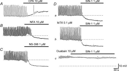
Figure 2. Pharmacological properties of spontaneous depolarizations in CCSM cells.
In a CCSM preparation, CPA (10 μm) depolarized the membrane by about 10 mV and prevented the generation of spontaneous action potentials (A). In a different preparation, niflumic acid (NFA, 10 μm) hyperpolarized the membrane by about 5 mV and abolished spontaneous action potentials (B). NS-398 (1 μm) hyperpolarized the membrane by about 10 mV and prevented action potential generation (C). In another preparation, SIN-1 (1 μm) hyperpolarized the membrane by about 10 mV and abolished action potentials (Da). In the same preparation which had been exposed to IbTX (0.1 μm), SIN-1 (1 μm) hyperpolarized the membrane by about 10 mV and prevented action potential generation (Db). In the presence of ouabain (10 μm), SIN-1 (1 μm) hyperpolarized the membrane by a few mV and failed to abolish membrane oscillations (Dc). The scale bar on the right of Dc applies to all traces. Resting membrane potentials were −48 mV for A, −45 mV for B, −46 mV for C and −48 mV for D.

