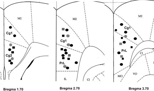
Figure 1. Location of neurones recorded in the anterior cingulate cortex (ACC).
Coronal sections from the caudal to rostral regions of the ACC (i.e. bregma 1.70, 2.70 and 3.70 mm) show the electrophysiological recording sites (adapted from the atlas of the rat brain by Paxinos & Watson (1998)). Squares and circles indicate ACC neurones in the control and EA rats, respectively. Black symbols represent CRD-excited neurones and grey symbols represent CRD-inhibited neurones. C1, claustrum; Cg1, cingulate cortex, area 1; Cg2, cingulate cortex, area 2; IL, intralimbic cortex; M2, secondary motor cortex; MO, medial orbital cortex; PrL, prelimbic cortex; VO, ventral orbital cortex.

