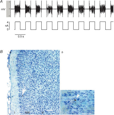
Figure 2. Juxtacellular labelling of an identified anterior cingulate cortex (ACC) neurone with neurobiotin.
A, Juxtacellular iontophoresis with positive current pulses (250 ms on/250 ms off, lower tracing shows current) of 2–4 nA produced bursts of action potentials that were continuously monitored and served as a measure of the magnitude of membrane disruption that resulted in cell labelling with neurobiotin. B, microphotograph of neurones in the ACC labelled with neurobiotin a, thionine-stained coronal section shows the laminar distribution of distension-excited neurones in the ACC. b, higher magnification of the neurobiotin-labelled pyramidal ACC neurone located in layer II/III in a (arrow). Scale bars: a, 250 μm; b, 50 μm.

