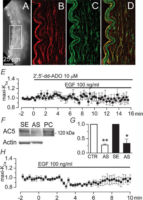
Figure 4. Adenylate cyclase 5 (AC-5) mediates maxi-KCa channel activation by EGFR.
A–D, fluorescence image of rat basilar artery immunolabelled for AC-5 (A and B) and coimmunolabelled for caveolin-1 (C); superimposed images of (B) and (C) demonstrate colocalization of AC-5 and caveolin-1 on plasmalemmal membranes of endothelial and smooth muscle cells (D); note autofluorescence of basal lamina at wavelengths for FITC filter (C), but not at wavelengths for CY3 filter (A and B). E, normalized change (mean ± s.e.m.) in membrane current measured using our standard conditions, after addition of EGF (100 ng ml−1) in the presence of 2′,5′-dd-ADO (10 μm) in the bath; 5 cells. F and G, Western blots (F) and bar graphs of densitometric analysis of Western blots (G) for AC-5 in basilar artery lysates from controls (CTR and SE) and from AC-5 knock-down (AS) rats; for AC 5-knock-down, rats underwent constant infusion of antisence (AS) oligodeoxynucleotide directed against AC-5 into the cisterna magna for 4 days (2 groups of 3 rats each); for controls, we used both normal, untreated rats (CTR; 3 rats), and rats with 4-day infusion into cisterna magna of corresponding sense (SE) oligodeoxynucleotide (3 rats); bar graphs show values normalized to corresponding controls, after first normalizing data from individual lanes to density of total actin; *P < 0.05; **P < 0.01; panel F also shows blots for total actin and a positive control (PC) for AC-5 (5 ng rat recombinant AC-5 obtained from FabGennix, Frisco TX). H, normalized change (mean ± s.e.m.) in membrane current in basilar artery VSMC from AC-5 knock-down rats, measured using our standard conditions, after addition of EGF (100 ng ml−1); 7 cells.

