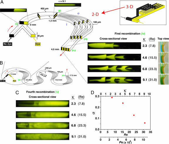
Fig. 2.
P-SAR micromixer incorporating four split streams. (A) Planar 2D microchannel geometry capable of generating alternating lamellae of individual fluid species in a split-and-recombine arrangement (400 μm wide; 29 μm tall; 630 μm radius of curvature; Re and κ computed based on the 400-μm wide segment; i and o denote the inner and outer channel walls, respectively). Flow schematics are shown inside the channel; corresponding confocal images are shown outside. Parallel streams of different species enter the curved microchannel (i) and experience a transverse flow generated by the counterrotating vortices above and below the channel midplane that induce a corresponding pair of 90° rotations in the fluid (ii). At this point (1.2 mm downstream from entrance), the flow is split into four parallel streams that proceed along curved trajectories inducing a second pair of 90° fluid rotations in each stream (between ii and iii). Alternating lamellae of the two species are generated when the streams are rejoined 4 mm downstream from the entrance (iv). Cross-sectional confocal images and the corresponding top-view images taken after the first recombination are shown. (Inset) Conventional 3D microchannel design required to achieve an equivalent lamination effect. (B) Schematic of a microchannel incorporating a series of successive P-SAR mixing elements. (C) Confocal cross-sectional images taken after the fourth recombination (position v in B). As κ is increased, the two species become almost completely intermixed, as indicated by uniform fluorescence over the channel cross-section. (D) Plot of σ computed from the confocal image sequence in C as a function of the Dean, Reynolds, and Péclet numbers (Pe was computed using D = 3 × 10⁻⁶ cm2/s for Rhodamine 6G).

