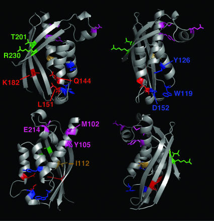
Fig. 1.
Amino acids changed in this study shown in the B. suis VirB8sp x-ray structure. Ribbon representation showing a model of the VirB8sp structure in different orientations. Dimer interface residues E214, M102, and Y105 are shown in magenta; active-site groove residues K182, L151, and Q144 are shown in red; β-sheet's solvent-exposed face residues R230 and T201 are shown in green; very high-identity patch Y126, D152, and W119 are shown in blue; and I112 is shown in brown. The model was generated with macpymol (pymol.sourceforge.net) based on the VirB8sp Protein Data Bank file, www.pdb.org (PDB ID code 2BHM).

