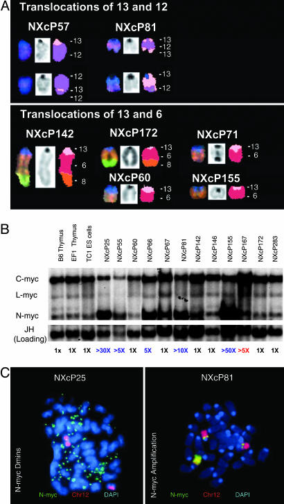
Fig. 3.
Recurrent translocations and amplifications in NXc/−p53−/− MBs. (A) Recurrent translocations involving chromosome 13 determined by SKY analyses of NXc/−p53−/− tumor metaphases. (A Upper) Clonal translocations involving chromosome 12 observed in 2 of 11 tumors. (A Lower) Clonal translocations involving chromosome 6. Translocations of the centromeric region of chromosome 13 onto the distal portion of the q arm of chromosome 6 were the most frequently observed in 5 of 11 tumors. Additional chromosome 13 and unique translocations are shown in Table 1. chr13, pink; chr12, purple; chr6, red; chr8, orange. (B) Southern blotting reveals the majority of NXc/−p53−/− tumors harbor amplified N-myc. Twelve NXc/−p53−/− tumors and control DNA were digested with EcoRI and probed with N-myc, c-myc, L-myc probes and then Ig heavy chain JH probe as loading control. Quantification of N-myc amplifications is shown in blue; c-myc amplification, in red, is labeled below. (C) Amplification of N-myc manifested as double minutes (Dmins) (NXcP25; Left) or as chromosomal amplifications (NXcP81; Right) detected by FISH with an N-myc BAC (in green) and chromosome 12 paint (in red). Unamplified N-myc is located at both chromosome 12 centromeric regions. In NXcP81, the amplified N-myc is located in another chromosomal location.

