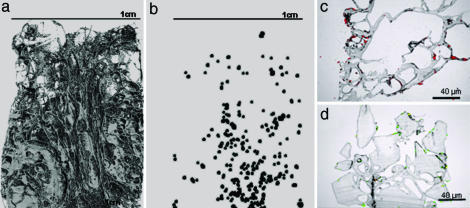
Fig. 3.
The distribution of mammalian cells after incorporation into PDLLA scaffolds by using a one-step scCO2 process. Location of cells throughout the scaffold observed by using μCT images of the polymer foam scaffolds (a) into which fixed cells were incorporated (b) (size and contrast increased for visualization). Cells were also observed throughout the scaffold in 10-μm sections highlighted with propidium iodide (c) and calcein acetoxymethyl ester and ethidium homodimer-1 (LIVE/DEAD) (d) prestained cells.

