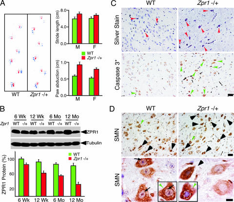
Fig. 1.
ZPR1 deficiency causes motor defects and apoptotic degeneration of facial motor neurons. (A) Abnormal gait of Zpr1+/− mice. Footprint patterns of 12-month-old wild-type (WT) and Zpr1+/− littermates (red, forepaws; blue, hindpaws) were examined. Quantitative analysis of stride length and paw abduction (26) for males (M) and females (F) was performed three times for each mouse (six mice per group). The results are presented as the mean ± SD. (B) Reduced Zpr1 gene dosage causes decreased expression of ZPR1 protein in the brain. Quantitation of immunoblots of ZPR1 expression in the brain was performed by using metamorph software (mean ± SD; four mice per group). (C) Low levels of ZPR1 cause apoptotic degeneration of facial motor neurons. Histochemical detection of facial motor neurons in sagittal sections of the brainstem from 12-month-old WT and Zpr1+/− mice. Brainstem section were examined by using silver stain to detect degenerating facial motor neurons (arrowheads) and counterstained with hematoxylin (Upper). Immunohistochemical detection of apoptosis used a monoclonal antibody to cleaved caspase 3 (arrowheads) in brainstem sections of 12-month-old mice (Lower). (Scale bar is 50 μm.) (D) ZPR1 deficiency causes degeneration of facial motor neurons. Immunohistochemical detection of SMN in sagittal sections of the brainstem from WT and Zpr1+/− mice. Degenerating neurons were found to be chromolytic (arrowheads) due to increased cytoplasmic accumulation of SMN. Neurons (green arrowheads) are also shown at higher magnification (Lower Inset). Arrows show the presence of prominent punctate intranuclear staining of SMN in WT neurons. (Scale bars, 50 μm in Upper and 10 μm in Lower.)

