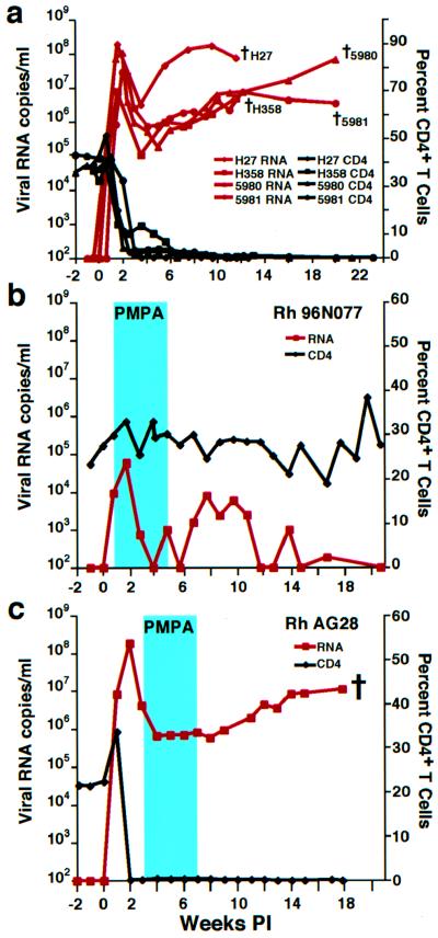
Figure 1.
SHIV and CD4+ T lymphocyte levels in infected rhesus monkeys. (a) Viral RNA loads (red) and CD4+ T cell levels (black) in rhesus macaques inoculated intravenously with 4.1 × 10⁵ (Rh H27 and Rh H358), 16,400 (Rh 5980), and 656 (Rh 5981) TCID50 of the SHIVDH12R stock virus are shown. The black crosses indicate the times of animal killing because of their deteriorating clinical condition. (b and c) Plasma viral RNA (red) and CD4+ T cell levels (black) in SHIVDH12R-infected rhesus macaques administered PMPA (30 mg per kg) for 28 days beginning 5 days (b) or 21 days (c) postinfection (PI) (blue rectangles). The cross indicates the time when monkey AG28 was killed.

