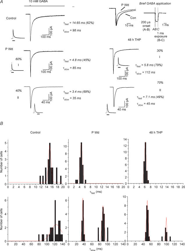
Figure 1. Progesterone withdrawal accelerates the deactivation of GABA-gated current.
A, representative traces showing responses to brief (∼ 1 ms) pulses of GABA (10 mm) recorded from outside-out patches of CA1 hippocampal pyramidal cells following progesterone withdrawal (P Wd), 48 h 3α,5β-THP (48 h THP) or sham conditions (Control). Each trace represents the average of 6–10 individual traces. (Fits are shown next to full traces.) The deactivation rate is best described as a biexponential decay, with a τfast in the range of 10–22 ms and a τslow of 80–145 ms for the control recordings. Following P withdrawal (P Wd), in Group I 60% of the current deactivated with a τfast of 3–6 ms (mean = 4.88 ± 0.61 ms), and a τslow of 80–120 ms (mean = 87.0 ± 12.0 ms). In Group II, 40% of the current recorded deactivated with a τfast of 3–7 ms, and a τslow of 30–40 ms. Forty-eight hours of treatment with 3α,5β-THP produced similar acceleration in deactivation times. Note that in both populations, τfast is significantly faster than control values, while in Group II τslow is also significantly faster than control. Average peak amplitude was unaffected by prior steroid treatment. The top trace indicates the open tip junctional current. (These results are representative of those recorded from 20 to 30 patches/group.) Inset: amplified traces illustrate an accelerated τfast following P Wd compared to control. Inset, representative open tip junction potential for a control recording. B, distribution of values for τfast and τslow for control (Con, left panels), progesterone withdrawal (P Wd, middle panels), and 48 h treatment with 3α,5β-THP (48 h THP, right panels). Values for τslow display a bimodal distribution for P Wd and 48 h THP conditions. All other distributions display a single mode.

