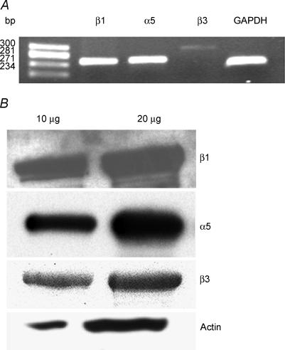
Figure 1. Expression of integrins in mouse papillary fibres.
A, RT-PCR analyses of α5, β1 and β3, integrins and GAPDH gene in papillary muscle of mouse hearts. Electrophoresis of PCR gene products on a 1.8% agarose gel were carried out and visualized with ethidium bromide staining. Molecular weight markers represent Hae III-digested φX-174 DNA. These data are representative of three independent experiments. B, representative Western blot analysis of α5, β1 and β3 integrins (n = 3). Ten and 20 μg of total heart homogenates were run on a 10% SDS-PAGE and probed with specific antibodies as detailed in Methods. The blots were re-probed with sarcomeric actin.

