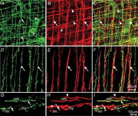
Figure 4. Relationship between inhibitory motor nerves and ICC-IM in the gastric corpus.
A, a confocal image reconstruction of nitric-oxide-synthase-like (nNOS-LI) nerve fibres within the circular, and longitudinal muscle layer of the corpus (green, double arrows) and cell bodies within the myenteric plexus region (*). B, ICC (red) within the same region of the corpus. ICC-IM in the circular and longitudinal layers, and ICC-MY at the myenteric plexus, are identified with arrowheads, arrows and *, respectively. C, double labelling of A and B, nNOS-LI nerve fibres are closely apposed to ICC-IM within the circular (arrowheads) and longitudinal (arrows) layers. D–F, nNOS-LI nerves (green, double arrows) and ICC-IM (red, arrows) within the longitudinal muscle layer. F, digital reconstruction of A and B showing the close apposition between nNOS nerves and ICC-IM (arrows) in the longitudinal layer. G–I, confocal images of cryostat sections through the corpus wall. G, nNOS-LI nerve fibres (double arrows). H, ICC-IM (arrowheads and arrows) and ICC-MY (*). I, digital reconstruction of panels G and H, confirming the close apposition of nNOS-LI and ICC-IM in the circular (arrowheads) and longitudinal muscle (arrows) layers. Scale bar in F is representative for panels A–F, and scale bar in I is representative for panels G–I. Digital reconstructions were A–C, 25 × 0.5 μm; D–F, 3 × 0.5 μm; G–I, 3 × 0.6 μm.

