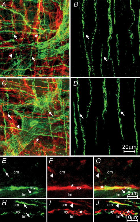
Figure 5. Relationship between excitatory and inhibitory motor nerves and ICC-IM in the longitudinal layer of the gastric antrum.
A, a confocal image reconstruction of a double-labelled image of vAChT-LI nerve fibres (green, double arrows) and ICC-IM within the circular (red, arrowhead) and myenteric plexus region (red, *) of the antrum. B, a double-labelled image of vAChT-LI and ICC within the longitudinal muscle layer of the same region of antrum. vAChT-LI nerve fibres (double arrows) but no ICC-IM were observed in the longitudinal layer of the antrum. C and D, a confocal reconstruction of a double-labelled image of nNOS-LI nerves (green, double arrows) and ICC-IM in the circular layer (red, arrowhead), and ICC-MY (red, *) in the myenteric plexus region. D, a double-labelled image of nNOS-LI nerve fibres (double arrow) within the longitudinal layer, and an absence of ICC-IM in this layer in the antrum. E–G, confocal reconstructions of cryostat sections revealing vAChT-LI nerve fibres (green, double arrows) in the circular (cm) and longitudinal (lm) layers (E), and ICC in the circular (ICC-IM, arrowhead) and myenteric plexus (ICC-MY, *) region (my) of the antrum, but not in the longitudinal muscle layer (F). Double labelling (G) shows vAChT-LI nerves in close apposition to ICC-IM in the circular (arrowhead) but not in the longitudinal layer (double arrow). H–J, confocal reconstructions of a transverse cryostat section of the antrum. nNOS-LI nerve fibres (green, double arrows) were observed in the circular (cm) and occasional fibres in the longitudinal (lm) layer. In I, ICC (red) were distributed similar to that described for F. J, a double-labelled image revealing the close apposition between nNOS-LI nerve fibres (double arrows) and ICC-IM (arrowhead) in the circular layer but not in the longitudinal layer of the antrum. Scale bar in D is representative for A–D. Scale bar in G is representative for E–G. Scale bar in J is representative for panels H–J. Digital reconstructions were: A, 15 × 1.0 μm; B, 4 × 1.0 μm; C, 25 × 0.6 μm; D, 3 × 0.6 μm; E–G, 4 × 0.5 μm; H–J, 3 × 0.6 μm.

