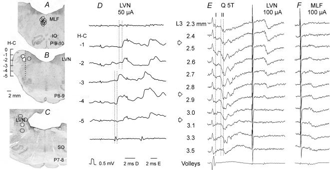
Figure 1. Reconstructions of the locations of the stimulating electrodes.
A–C, locations of the stimulating electrode tips, as defined by the electrolytic lesions made at the end of the experiments, in the ipsilateral medial longitudinal fascicle (MLF) in planes P9–10 and in the ipsilateral lateral vestibular nucleus (LVN) in Horsley-Clarke's planes 8–9 and P7–8, respectively, These are superimposed on representative sections of the brainstem, cut in the plane of the electrode insertions (at the angle of 30 deg). IO, inferior olive; SO, superior olive. D, monosynaptic EPSPs evoked in one of the interneurones in the same experiment as in E, by stimuli applied at different depths at Horsley-Clarke's (H-C) coordinates H0 to H-5 along the electrode track indicated in B and descending volleys from the depth −2 (top trace) and −4 (bottom trace). The two dotted lines indicate the descending volleys and the onset of the EPSPs evoked from the dorsal and ventral stimulation sites (at latencies 0.7 and 0.5 ms from the volleys, respectively). E, field potentials recorded at different depths from the surface of the spinal cord (indicated to the left) along an electrode track crossing grey matter in the L3 segment of the spinal cord at an angle of 10 deg (tip directed lateral). These were evoked by stimulation of the quadriceps nerve (Q) at 5 times threshold (T) and from LVN. The three dotted lines indicate the onset of monosynaptic field potentials from group I afferents and from group II afferents at most dorsal locations and at the depths of 3.3 and 3.5 mm. F, field potentials along another electrode track in the same experiment, 200 μm more medial. Arrows indicate the levels of the maximal field potentials from the LVN, from group II afferents in lamina VIII and from MLF within which interneurones with input from these sources were searched for. In this and the following figures the negativity in the microelectrode (intracellular or extracellular recordings) is downwards and in records from the surface of the spinal cord upwards.

