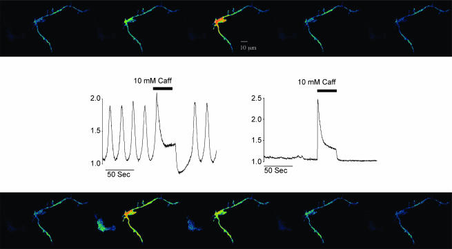
Figure 1. Ca2+ changes in interstitial and smooth muscle cells.
Sequence of 10 frames before (top panel, taken at 4 s intervals) and during addition of 10 mm caffeine (bottom panel, at 3 s intervals). The highly branched cell on the right of each frame is a typical interstitial cell, while the darker spindle-shaped cell on the left, scarcely visible under control conditions, is a smooth muscle cell. Regular spontaneous increases in fluorescent intensity were apparent in the perinuclear region of the branched cell, while the smooth muscle cell remained dark. However, when 10 mm caffeine was added both cells lit up simultaneously (frame taken at 64 s). When the caffeine-induced transient was complete, the smooth muscle cell again became dark, and remained so for the rest of the experiment, while the interstitial cell, after a brief pause, resumed its oscillatory behaviour. This experiment also illustrated the different contractile properties of both cells. Thus, while the smooth muscle cell contracted vigorously in response to caffeine addition, the interstitial cell did not contract either in response to the spontaneous increases in intracellular Ca2+ or in response to the caffeine-induced Ca2+ transient. Scale bar, 10 μm.

