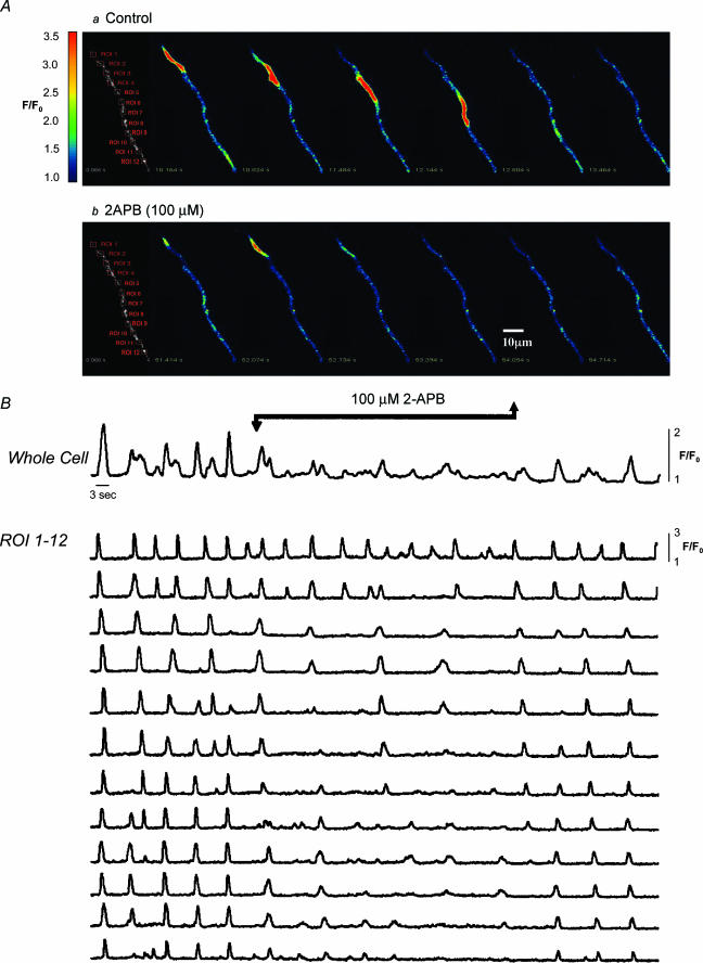
Figure 3. Effect of 2-APB on wave propagation.
Series of images of the initiation and spread of a Ca2+ wave under control conditions (A, upper panel) The wave arose at the top of the cell (region of interest (ROI) 1) and propagated without decrement to the other end of the cell. Occasionally the wave originated at ROI 9 and propagated in the opposite direction, but generally the global event was well coordinated throughout the cell (as is apparent from the plotted regions of interest in B). In the presence of 2-APB (A, lower panel) the pattern was quite different. Oscillations continued in all parts of the cell, but these were now propagated to only one or two of the neighbouring ROIs, with the result that activity in the cell as a whole was now very poorly coordinated. Upon washout of 2-APB, the well-coordinated global events were quickly restored. Scale bar, 10 μm.

