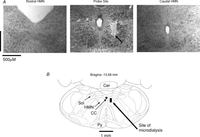
Figure 1. Example showing location of microdialysis probe.
A, example of a lesion site made by the microdialysis probe in the hypoglossal motor nucleus (HMN). The arrow shows the location of the lesion site in the HMN. Sections both rostral and caudal to the probe site are also shown. B, schematic representation of the lesion site from the histological section. The size of the bar represents the apparent size of the lesion. Abbreviations: Cer, cerebellum; Sol, nucleus tractus solitarius; Py, pyramidal tract; CC, central canal.

