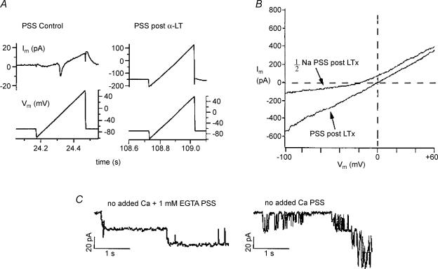
Figure 7. Ionic dependence of currents induced by α-LT in voltage-clamped canine β-cells.
A, compares the Im–Vm relationship of a cell before versus after application of 5 nm α-LT in 2 mm Ca2+ PSS. The cell was patch clamped with a pipette filled with standard K+-IS. In each case Im was recorded in response to 250-ms voltage ramps from −100 mV to +60 mV (details in the text). B, effect of [Na+]o reduction on the Im–Vm relationship of the toxin-treated cell. When half of the extracellular [NaCl] was replaced with N-methyl-d-glucamine (NMDG) chloride, the zero current potential of the cell is shifted negatively by 15 mV and the Im–Vm relationship shows outward rectification (1/2Na PSS, post α-LT), as compared with the that recorded in 2 mm Ca2+ (PSS, post α-LT). C, compares single-channel currents recorded from a toxin-treated cell bathed in no added Ca2+ PSS (right trace) with those from a toxin-treated cell bathed in no added Ca2+/EGTA PSS (left trace).

