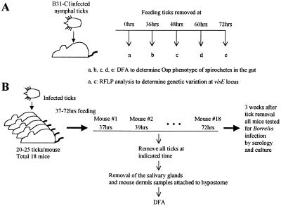
Figure 1.
Outline of experimental approach. (A) B. burgdorferi Osp phenotype in the tick gut. Infected ticks were placed on mice and removed at the indicated times. Twelve unfed and 93 fed nymphal ticks (12 ticks from 36 hr, 39 ticks from 48 hr, 36 ticks from 60 hr, and 6 ticks from 72 hr) were removed. The guts were dissected and examined by DFA to determine the OspA and OspC phenotype within the gut. DNA was purified from gut homogenates prepared from unfed and partially engorged (48-hr) ticks to determine the extent of variation at the B. burgdorferi vlsE locus. (B) B. burgdorferi infection of tick salivary glands and mouse dermis. A total of 18 mice were infested with 20–25 infected nymphal ticks. All of the ticks were removed from individual mice at 2-hr intervals starting at 37 hr and ending at 72 hr into the blood meal. Mouse and tick samples were analyzed for B. burgdorferi infection and Osp phenotypes.

