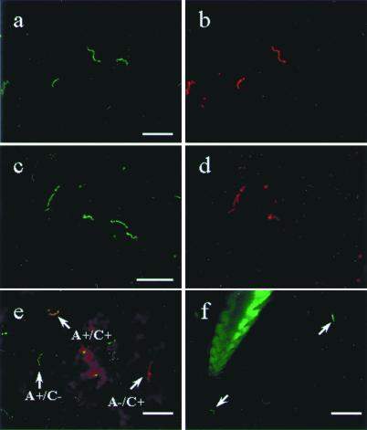
Figure 4.
Direct dual immunofluorescence of B. burgdorferi in the gut of partially engorged (48 hr) ticks and in the mouse dermis. (a and b) The same field double-labeled with a FITC-conjugated polyclonal B. burgdorferi antibody (a) and a TR-conjugated mAb against OspA (b). (c and d) The same field labeled with FITC-conjugated polyclonal Borrelia antibody (c) and a TR-conjugated mAb against OspC (d). (e) Merged image of spirochetes stained with Alexa 488-conjugated OspA (green) and TR-conjugated OspC (red). Three phenotypes were observed in e: arrows, Borrelia producing only OspA in green, only OspC in red, and both OspA and OspC in orange. (f) Mouse skin sample attached to the hypostome of a tick that had fed for 40 hr is labeled with the FITC-conjugated polyclonal Borrelia antibody. The hypostome itself autofluoresces, and bacteria staining with the antibody are indicated by the arrowhead. (Bars in a, c, and e represent 20 μm, and the bar in f represents 50 μm.)

