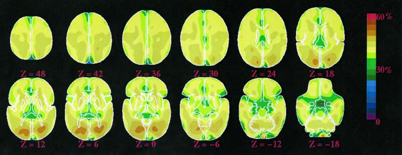
Figure 4.
Maps of the fraction of oxygen extracted by the brain from arterial blood (oxygen extraction fraction or OEF expressed as a percentage of the available oxygen delivered to the brain). The data come from 19 normal adults (group I, Table 1) resting quietly but awake with their eyes closed. The data were obtained with PET. Despite an almost 4-fold difference in blood flow and oxygen consumption between gray and white matter, the OEF is relatively uniform, emphasizing the close matching of blood flow and oxygen consumption in the resting, awake brain. Areas of increased OEF can be seen in the occipital regions bilaterally (see text for discussion).

