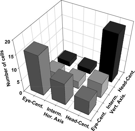
Figure 2. Distribution of spatial reference frames.
As described in more detail in the main text we determined for each individual neurone its reference frame for visual spatial information along the horizontal and vertical axis (see Fig. 1). Neurones whose visual RF shifted completely with the eye were considered eye-centred. Those neurones whose visual RF did not shift at all with the eye were considered head-centred. Interestingly, a number of neurones fell in an intermediate class that is neither eye- nor head-centred.

