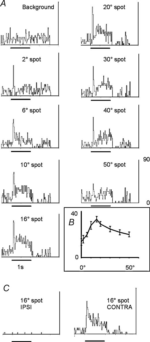
Figure 2. PSTHs representing a ‘classic’ monocular receptive field.
A, PSTHs showing the responses of an exclusively contralaterally driven cell to flashed spots of light of increasing diameter. Bin size 25 ms. This cell showed an initial transient response, followed by a more sustained component, most obvious with larger stimulus sizes. Peak responses were seen to stimuli of the order of 16–20 deg in diameter. This is most evident in the inset (B), a histogram of the response magnitudes to the various stimulus sizes. Values are the total number of spikes fired ± 1 s.e.m.C, activity of this cell during visual stimulation via the ipsilateral eye, left, and repeated contralateral stimulation using a 16 deg diameter stimulus, right.

