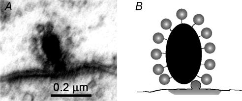
Figure 2. The ribbon synapse.
A, transmission electron micrograph of a synaptic ribbon of an inner (cochlear) hair cell in a 2-month-old rat. The electron-dense ribbon is surrounded by a halo of small vesicles (∼30 nm diameter). The plasma membranes are thickened beneath the ribbon, that of the postsynaptic afferent neurone more obviously so. (Micrograph by T. Pongstaphone, unpublished.) B, ribbon schematic showing vesicles tethered to the dense body (ribbon), with one vesicle having fused to plasma membrane to release its contents (grey ‘cloud’ flattened by adjoining postsynaptic membrane).

