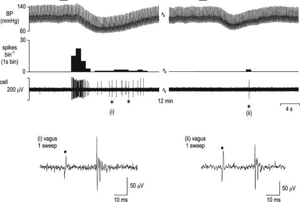
Figure 1. Identification of nucleus tractus solitarius neurones receiving vagal cardiopulmonary receptor inputs.
The response of a single nucleus tractus solitarius (NTS) neurone to activation of cardiopulmonary afferents by intra-atrial administration of phenylbiguanide (PBG, 12 μg kg−1, 20 μl) at the horizontal bar is shown in the left panel. This response is mediated by vagal afferents as the response to the same stimulus is abolished following bilateral cervical vagotomy (right panels). The neuronal recording is still intact following vagotomy since electrical stimulation of the cervical vagus central to the vagotomy (⋆) still evokes activity in the neurone (right panel). Top panels – from top, traces show arterial blood pressure (BP; mmHg), a continuous rate histogram of neuronal activity (spikes bin−1) and the raw recording of neuronal activity (μV). Bottom panels, single sweeps of neuronal activity evoked by electrical stimulation of the vagus nerve (•) before (i) and following (ii) vagotomy distal to the stimulating electrode at the points marked ⋆ in the panels above.

