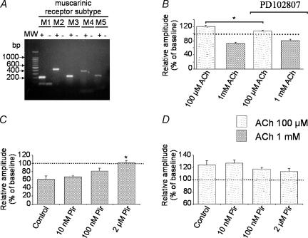
Figure 3. Effects of muscarinic receptor antagonists on ACh modulation of synaptic transmission.
Pharmacological antagonists of mAChRs were bath applied in visual cortex slices of SLJ–C57BL/6J mice. A, agarose gel electrophoresis showing the products from reverse transcription and PCR of total RNA from visual cortex of SLJ–C57BL/6J wild-type mice using M1, M2, M3, M4, and M5 mAChR gene-specific primers. Amplification of cDNA from reverse transcription reactions is indicated as +. Control reactions to check for contamination with genomic DNA were run without reverse transcribing the RNA samples (indicated with –). All mAChR genes are expressed in the visual cortex of control mice. B, in the presence of the selective M4 mAChR antagonist PD102807 (0.5–1 μm), there was a significant reduction of 100 μm ACh-induced (n = 13) facilitation without affecting 1 mm ACh-induced depression in FP amplitudes (n = 7).(C and D). The mAChR antagonist pirenzepine was bath-applied at three different concentrations (10 nm, 100 nm, and 2 μm). Application of pirenzepine for 10 min did not modify basal FP amplitudes (data not shown). C, depression of FPs induced by 1 mm ACh was prevented by 2 μm but not by 10 and 100 nm pirenzepine. *P < 0.01; other conventions as in Fig. 2. D, pirenzepine (Pir) at the concentrations used did not significantly change the facilitation of FP amplitudes induced by 100 μm ACh.

