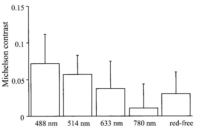
Fig. 1.

Michelson contrasts for internal limiting membrane peeling for idiopathic macular hole in scanning laser ophthalmoscope images at 488 nm, 514 nm, 633 nm, and 780 nm and red-free fundus photograph (red-free). Michelson contrasts in scanning laser ophthalmoscope images at 488 nm and 514 nm were significantly larger than those in other images.
