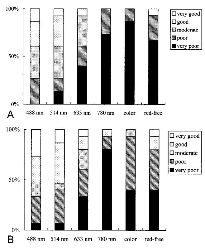
Fig. 3.

Results of grading the margin of internal limiting membrane peeling (A by T.A. and B by K.Y.). The bar graphs show results for each grader for each image type: scanning laser ophthalmoscope images at 488 nm, 514 nm, 633 nm, and 780 nm; color fundus photograph (color); and red-free fundus photograph (red-free). The scanning laser ophthalmoscope images at 488 nm and 514 nm were graded superior to other images.
