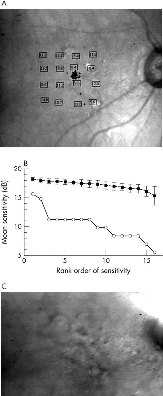
Figure 4.

Blue on yellow perimetry results for an 86 year old female patient with visual acuity (VA) 20/25, good fixation, and distinct soft drusen. (A) Results in dB, plotted on the final confocal infrared image. This is the only patient with atrophic areas. One is beneath the bottom, rightmost stimulus loci. The other is in the top row, between two loci. The worst sensitivity for this patient was at the atrophic site. Other loci with poor sensitivity, near the optic nerve head, fell onto regions of confluent material and hyperpigmentation (see C). (B) Bebié function for the patient v the average of controls’ data, as in figure 3B, showing most loci had worse sensitivity than the controls’ data, as well as a greater difference among loci in sensitivity as seen from the slope of −0.051. (C) Indirect mode infrared image showing multiple soft drusen in a 25×19 degree area. There is a region of image saturation over the optic nerve head. There is a thickened area of confluent material near the optic nerve head, not to be mistaken for atrophy but containing clumped hyperpigmentation.
