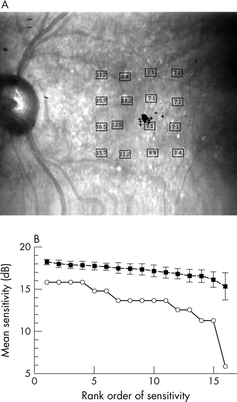
Figure 5.

Blue on yellow perimetry results for an 85 year old male patient with visual acuity (VA) 20/25, good fixation, and confluent soft drusen. (A) Results in dB, plotted on the final confocal infrared image. (B) Bebié function as in figure 3B with a slope of 0.031, showing that most loci had worse sensitivity, as well as a somewhat greater difference among loci in sensitivity.
