
Fig. 1.
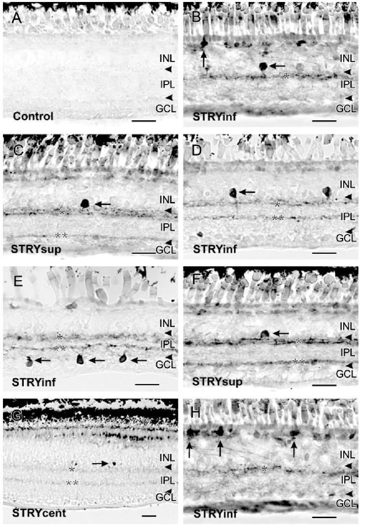
A: Control retinas had no cGMP-LI. B: In the inferior retina below the visual streak(STRYinf), 100 μM strychnine increased cGMP-LI in sGC A1 amacrine cells (horizontal arrow) and sGC B2 bipolar cells (vertical arrow). A single band of cGMP-LI was present in S20 of the IPL (single asterisk) in the inferior retina close to the ora serrata. C: In the superior retina above the visual streak (STRYsup),100 μM strychnine increased cGMP-LI in sGC A2 amacrine cells (horizontal arrow). Two labeled bands were present at S15-30 and S80 of the IPL (single and double asterisks). D: Labeled sGC A3 amacrine cells were seen in the inferior retina close to the visual streak (horizontal arrow). Two bands at S20 and S60 of the IPL were also visible (single and double asterisks). E: Labeled sGC 4 cells were seen in the GCL of the inferior retina (horizontal arrows). F: Labeled A sGC A11 cells were seen in the superior retina (horizontal arrow). G: In the central retina(STRYcent), 100 μM strychnine stimulated increases in cGMP-LI in two faint bands at S20 and S65 of the IPL. Some GC A1-like amacrine cell somata were also seen (horizontal arrow). H: More bipolar cells with strong cGMP-LI were seen in the inferior retina (vertical arrows). Scale bars = 25 μm.