
Fig. 5.
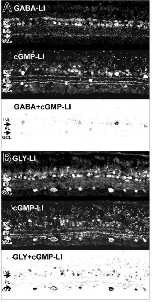
Colocalization of cGMP with GABA or GLY. A: The top panel shows strong GABA-LI in select amacrine cell somata, and in synaptic boutons in the IPL. The middle panel shows strong cGMP-LI in a retina treated with NO donor. The bottom panel shows a contrast inverted image of the colocalization of GABA-LI and cGMP-LI in some amacrine cell somata and in some processes and synaptic boutons in the IPL. B: The top panel shows strong GLY-LI in amacrine cell somata, somata in the GCL, and in synaptic boutons in the IPL. The middle panel shows strong cGMP-LI in a retina treated with NO donor. The bottom panel shows a contrast inverted image of the colocalization of GLY-LI and cGMP-LI in numerous amacrine cell somata, somata in the GCL, and in processes and synaptic boutons in the IPL. These data were the average of 30 images from 3 animals (10 per animal) for each antibody.