
Fig. 13.
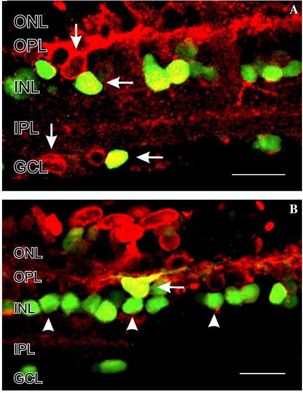
Colocalization of NO-IF and nNOS-LI in salamander retinal slices stimulated with 50 μM nicotine. In these images the NO-IF is green, the nNOS-LI is red, and yellow indicates that it was nNOS that was producing the NO-IF. The yellow double labeling indicates that nicotine primarily activates nNOS in somata in the INL and GCL (A and B, horizontal arrows). (A) In some horizontal cells in the INL and in some somata in the GCL, the nNOS is not activated by nicotine (vertical arrows). In some somata in the INL there is NO-IF but no nNOS-LI, which potentially indicates the activation of other isoforms of NOS than nNOS. Scale bars, 20 μm.