
Fig. 4.
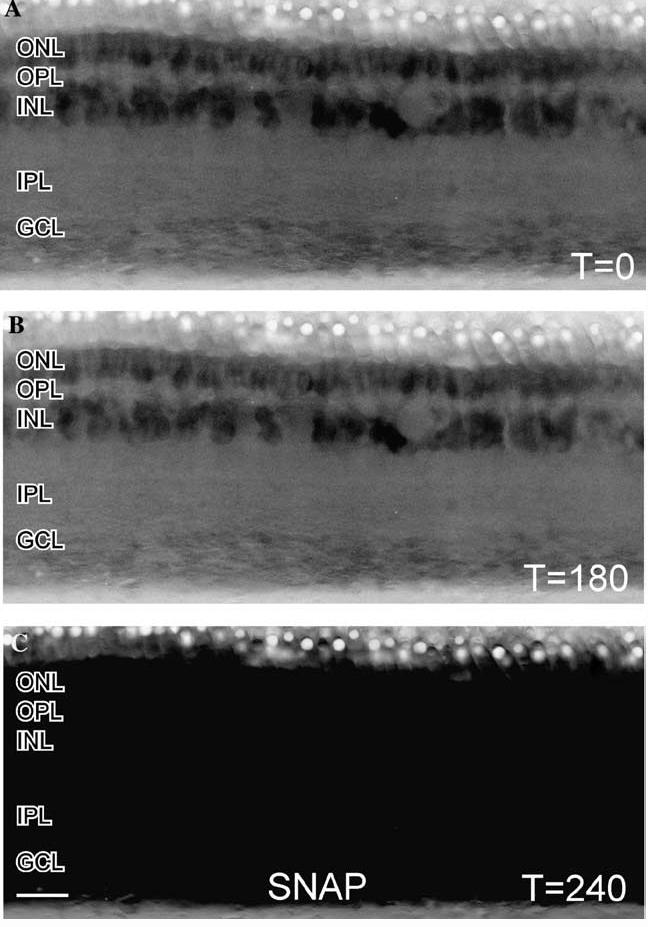
Effects of NO-donor on a turtle retinal slice loaded with DAF. (A) In these contt reversed images of a retinal slice loaded with DAF and illuminated with 488 nm light, there was some faint NO-IF apparent in the ONL and INL. (B) After 180 s of illumination there was only a slightly perceptible decrease in the levels of NO-IF which indicates that DAF is not activated by light alone. (C) Stimulation with 100 μM of the NO-donor SNAP at 180 s caused a dramatic and uniform increase in NO-IF throughout the slice by 240 s. This broad increase in NO-IF indicated that the entire slice was uniformly loaded with DAF. Scale bar, 20 μm