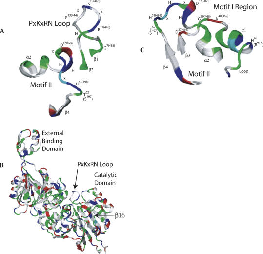
FIGURE 2.
Views of tRNase Z taken from the B. subtilis structure (de la Sierra-Gallay et al. 2005; PDB ID 1Y44). Motif II and the other active site residues (Motifs III–V) are buried in the catalytic domain of the A subunit, and the PxKxRN loop appears to cover the entrance to the active site. (A) PxKxRN loop and Motif II. (B) entire Bsu. tRNase Z structure model in the same front view as in A with the external binding domain in the upper left. (C) Motif I region and Motif II; the best view was obtained by rotating ∼180° so that the external binding domain is in the upper right (a back view). The amino acid, α-helix, and β-sheet nomenclature refer to the B. subtilis structure. Residue numbers in parentheses refer to the fruit fly protein.

