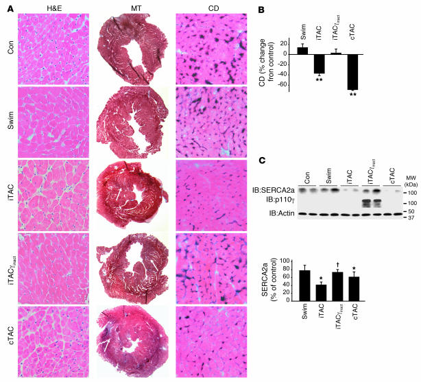
Figure 6. Preservation of βAR signaling through targeted PI3K inhibition is associated with preservation of capillary density and SERCA2a levels.
(A) Representative staining of cardiac sections with H&E (magnification, ×400), MT (magnification, ×20), and endothelial alkaline phosphatase (to determine capillary density; magnification, ×400) after 1 week of training. (B) Quantification of capillary density in cardiac sections stained for endothelial alkaline phosphatase expressed as percent reduction from control (n = 4–6 hearts per group). **P < 0.01 versus control, swimming, and iTACγinact; Student’s t test with Bonferroni correction. (C) Immunoblotting analysis of SERCA2a, actin, and PI3Kγinact protein levels in hearts from experimental design II. Densitometric quantification of SERCA2a levels is shown in the bottom panel (n = 6–10 per group). *P < 0.05 versus control, †P < 0.05 versus iTAC, ANOVA with Neuman-Keuls correction.

