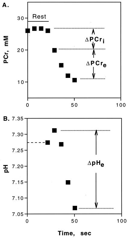
Figure 1.
PCr (A) and pH (B) levels starting at the onset of ischemia in resting muscle and through ischemic rattling. The contractile ATP demand is measured by Δ[PCr]i. After the onset of acidosis, the changes in [PCr] (Δ[PCr]e) and pH (ΔpHe) reflect glycolytic H+ production (Eq. 1; ref. 14). Values are means, n = 6. The dashed line in B is the resting muscle pH from high-resolution spectra.

