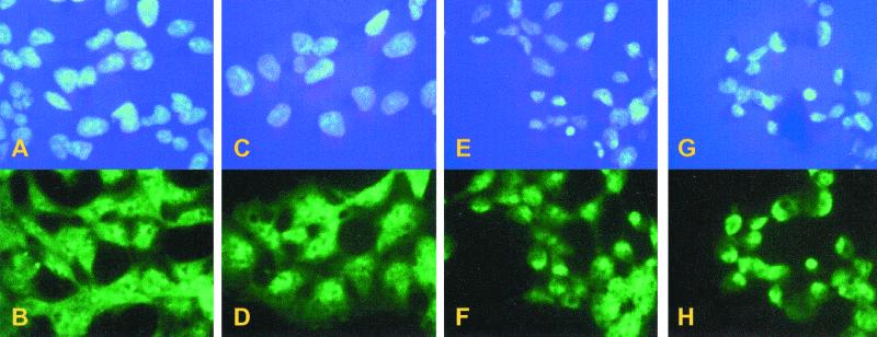
Figure 5.
In situ hybridization of mRNA in FSN cells. Cells were not induced (A and B) or induced for 4 h (C and D), 8 h (E and F), and 16 h (G and H). Cell nuclei were stained by using DAPI (Upper). mRNA was hybridized with a digoxigenin-coupled oligo(dT) and then visualized by using FITC-conjugated anti-digoxigenin Fab fragments.

