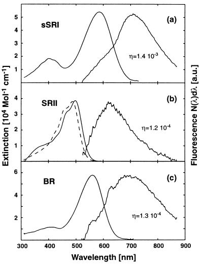
Figure 1.
Absorption and fluorescence spectra of sSRI (a), sSRII (b, dashed line), pSRII (b, solid line), and BR (c). Fluorescence spectra are scaled to the same height as the corresponding absorption spectra. A fluorescence spectrum of sSRII could not be recorded because of dominating buffer fluorescence around 560 nm. Buffer fluorescence also is visible in the short wavelength wing of the sSRI fluorescence band (a). In the fluorescence spectrum of BR the narrow shoulders at 560 and 620 nm arise from Raman scattering of water.

