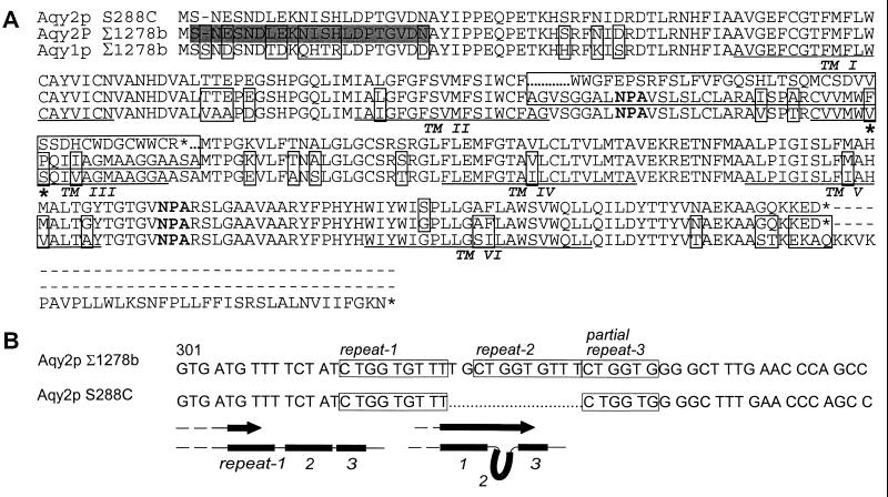
Figure 1.
Sequence alignment of Aqy1p and Aqy2p. (A) Comparison of Aqy1p from Σ1278b, Aqy2p from S288C (database strain), and Aqy2p from Σ1278b. Residues that differ between sequences are boxed. The deletion in S288C AQY2 is marked with dots. Aqy2p residues 141 and 142 are identified with asterisks. Putative transmembrane domains are identified (TM1–6). The shaded residues correspond to the sequence of the peptide used to make anti-Aqy2p antibodies. (B) The deletion in S288C and Σ1278b AQY2 with nucleotide repeats is boxed. Schematic diagram shows a possible mechanism for repeat removal that could explain the S288C deletion.

