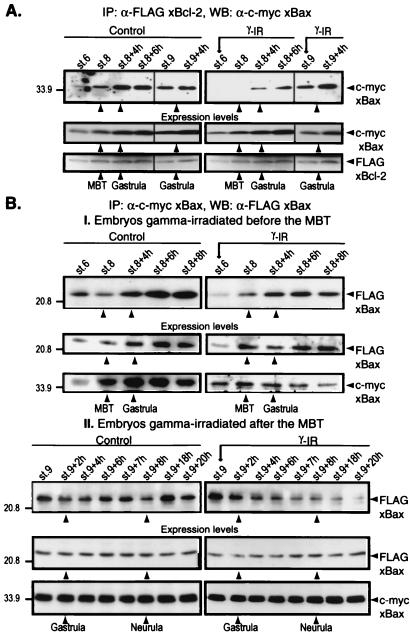
Figure 2.
(A) Interaction between xBcl-2 and xBax changes at the MBT. Embryos were injected at the one-cell stage with a mixture of FLAG-tagged xBcl-2 and c-myc-tagged xBax mRNAs. The embryos were not irradiated (control) or were irradiated 3 h (stage 6) or 7 h (stage 9) after fertilization, collected at different times, and frozen. Samples equivalent to five embryos were immunoprecipitated with anti-FLAG M2-agarose beads, and the immunocomplexes were subjected to Western blot analysis with anti-c-myc antibody. (B) Analysis of xBax homodimerization. Embryos were injected at the one-cell stage with FLAG-tagged and c-myc-tagged xBax mRNAs, were not irradiated (control) or were irradiated at either stage 6 (I) or stage 9 (II), and were collected at different times. Samples equivalent to five embryos were immunoprecipitated with anti-c-myc-agarose beads, and the immunocomplexes were blotted with anti-FLAG M2 antibody (I, Top, and II, Top). Total FLAG-xBcl-2, FLAG-xBax, and c-myc-xBax expression levels were assessed at all stages by Western blotting with specific anti-tag antibodies (A and B, I and II, Middle and Bottom). Arrows on the right denote the FLAG-tagged or c-myc-tagged protein.

