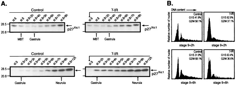
Figure 3.
(A) Analysis of the level of p27Xic1 in embryos irradiated either pre- or post-MBT. Embryos were not irradiated (control) or were irradiated (γ-IR) at the indicated stages, and samples were collected at the indicated times and analyzed by Western blotting with anti-p27Xic1 antibody. One embryo equivalent was loaded per lane. (B) Cell cycle profile of irradiated embryos. Embryos were irradiated at stage 9 and collected 2 and 6 h after irradiation. Embryonic nuclei were prepared from a single sibling group and analyzed by flow cytometry as described in Materials and Methods. Stages of the cell cycle are defined by DNA content, as measured by fluorescence intensity.

