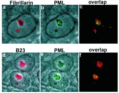
Figure 2.
Nucleolar localization of PML in MG132-treated MCF-7 cells. High magnification image of double staining for fibrillarin and PML or for B23 and PML. (a) Phase-contrast field combined with fibrillarin staining (red). (b) Phase-contrast and nucleolar PML (green) staining representing the same field as shown in a. (c) Overlap between fibrillarin and PML staining. (d) Phase-contrast field combined with nucleolar B23 staining (red). Panels e [phase-contrast and nucleolar PML staining (green)] and f overlap between B23 and PML.

