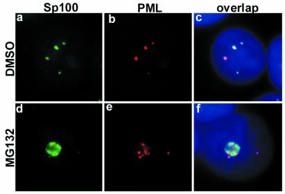
Figure 3.
Sp100 changes subcellular distribution and disassociates from PML upon MG132 treatment of MCF-7 cells. High magnification of Sp100 (green) and PML (red) double-staining of DMSO (a–c) or MG132-treated MCF-7 cells (d–f). (c and f) Overlap of Sp100 and PML. In DMSO-treated cells, Sp100 colocalized to a large extent with PML (c). After MG132 treatment, Sp100 completely changed its localization (d and f); it accumulated in the nucleoli along with PML but without any colocalization (f). DNA staining in blue.

