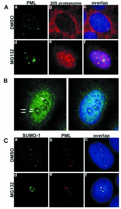
Figure 4.
(A) Double staining of PML (green) and proteasomes (red) in DMSO- or MG132-treated MCF-7 cells. In DMSO-treated cells, proteasomes were homogeneously distributed throughout the nucleus excluding nucleoli (b). Double staining showed no obvious colocalization between the two proteins (c). MG132 treatment changed the nuclear distribution of both PML and proteasomes; they both accumulated in the nucleoli as shown in panel (d–f). (B) Subnuclear localization of proteasomes in MG132-treated MCF-7 cells. Proteasomes (green) accumulate in the euchromatin areas and nucleoli and avoid peripheral (solid arrowheads) and perinucleolar (concave arrowheads) heterochromatin. (C) a–c represent double staining for SUMO-1 (green) and PML (red) in DMSO-treated MCF-7 cells. Overlap image shows complete colocalization of the two proteins (c). Upon MG132 treatment, SUMO-1 and PML accumulated in the nucleoli (d and e) without colocalization. DNA staining in blue.

