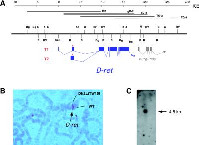
Figure 1.
The D-ret gene and transcripts. (A) Genomic organization of D-ret. The two alternatively spliced transcripts, T1 and T2, are schematically diagramed below the restriction map. Red triangle, translation initiator codon; An, poly(A). A nearby gene, burgundy, is also indicated. Shown above the restriction map are the genomic phage clones analyzed. (B) Heteroduplex loop formed between wild-type and Df(2L)TW161 chromosomes. D-ret (brown signal) is located at 39B02–39C02. (C) Northern blot analysis with 4 μg of poly(A)+ RNA from 0- to 24-h embryos. D-ret mRNA is detected as a 4.8-kb band.

