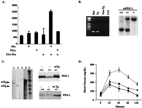
Figure 1.
Expression of wild-type or PBX interaction defective PDX-1 transgenes rescues glucose homeostasis in pdx-1 (+/−) mice. (A) Transient transfection assay of wild-type and PBX interaction defective (Pdxμ) PDX-1 constructs in 293T cells, with a TSEII luciferase reporter containing two PDX:PBX heterodimer recognition sites. Cells were transfected with wild-type or mutant PDX plus E2A-PBX expression vector where indicated. The first bar on the left represents control transfection with empty vector alone plus TSEII reporter. Luciferase activity shown after normalizing to β-galactosidase activity from cotransfected RSV-βgal plasmid. (B) Genotypic analysis of mice expressing PDX-1 transgene. (Left) Inheritance of PDX-1 transgene was evaluated by PCR assay with primers that are selective for rat PDX-1. (Right) Mice heterozygous or homozygous for targeted disruption of the murine PDX-1 gene were identified by genomic Southern blot analysis with a 32P-labeled PDX-1 cDNA probe. Insertion of the inactivating β-galactosidase gene is indicated by a 3.8-kb vs. a 3.0-kb EcoRI fragment. (C) Wild-type (Lane 1) and mutant (Lane 2) PDX-1 transgenes are comparably expressed in transgenic mice. (Left) RNase protection assay of total RNA from whole pancreas of adult mice, with the use of 32P-labeled wild-type PDX-1 antisense RNA probe. The shorter fragment in mutant mice results from RNase digestion in sequences encoding mutant FPWMK/AAGGQ motif. Lane 3, control nontransgenic littermate RNA; the rat PDX-1 probe does not recognize murine PDX-1. Lane 4, position of undigested PDX-1 antisense probe. Lane 5, molecular weight marker. (Right) Western blot assay of whole pancreas extract, showing levels of PDX-1 protein in wild-type, pdx-1(+/−), pdx-1(+/−);mTg, and pdx-1(−/−);wtTg mice. (D) Line graph of blood glucose levels in wild-type (—■—), pdx-1(+/−) heterozygous (– –⧫– –), pdx-1(+/−); wtTg, and pdx-1 (+/−);mTg mice (⋅⋅⋅●⋅⋅⋅) after i.p. glucose injection. Results for pdx-1(+/−); wtTg and pdx-1(+/−);mTg mice were indistinguishable.

