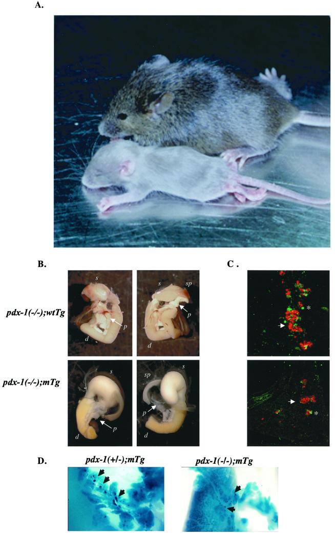
Figure 2.
Islet hypoplasia in pdx-1(−/−);mTg neonates. (A) Comparison of pdx-1(−/−);mTg (smaller) and wild-type (larger) littermates 16 days after birth. (B) Whole mount of pdx-1(−/−); wtTg and pdx-1(−/−); mTg mice. Ventral (Left) and dorsal (Right) views are included. p, pancreas; d, duodenum; sp, spleen; s, stomach. (C) Immunocytochemical analysis of insulin (red, arrows) and glucagon + somatostatin (green, asterisks) cells in sections from pdx-1(−/−); wtTg (Upper) and pdx-1(−/−):mTg (Lower). (D) Whole mount showing smaller developing islets (dark blue; arrowheads) in pdx-1(−/−) mTg compared with pdx-1(+/−) mTgt mice. Blue color indicates expression of β-galactosidase from the endogenous pdx-1 allele pdx-1lacZKO (1).

