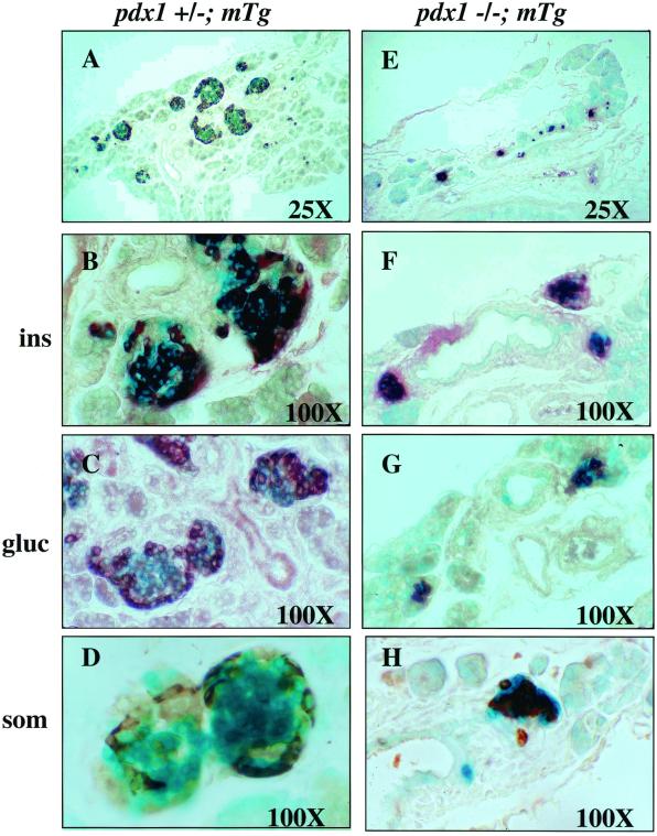
Figure 3.
Pancreatic islets of neonatal pdx-1(−/−);mTg mice are smaller and have disrupted architecture. (A–D) Sections of pdx-1(+/−); mTg islets. (E–H) Sections of pdx-1(−/−); mTg islets. pdx-1(−/−); mTg islets are much smaller and are located in close proximity to the ducts. Immunohistochemistry (brown) shows insulin-positive (F), glucagon-positive (G), and somatostatin-positive (H) cells in pdx-1(−/−); mTg islets. Glucagon- and somatostatin-positive cells (G and H) are not localized at the islet periphery as in wild-type islets (compare with C and D).

